
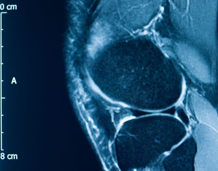
While in most patients, plain X-rays of knee joints are sufficient to evaluate the degree of osteoarthritis, sometimes MRI of the knee before genicular artery embolization “GAE” would be performed to evaluate the underlying cause of the knee pain, such as osteoarthritis, meniscal tears, ligament injuries, or other structural abnormalities. This imaging helps the healthcare provider assess the suitability of the patient for GAE and plan the procedure accordingly. Additionally, it provides valuable information about the anatomy of the knee and the distribution of blood vessels, which can be important for guiding the embolization procedure.
Quick Answer: Most patients considered for genicular artery embolization (GAE) only need standing knee X-rays to confirm and grade osteoarthritis. A knee MRI is added when the picture is unclear: when X-rays look mild but pain is severe, when there are locking or giving-way symptoms, or when another cause such as a meniscal tear, bone stress injury, osteonecrosis or a mass needs to be ruled out. The MRI helps confirm that inflammation-driven arthritis pain is the real target, so GAE is offered to the patients most likely to benefit.


Meet your vein specialist
Dr. Syed Raza, MD
Vascular & Interventional Radiology · Founder, Premier Vein & Vascular Center
Fellowship-trained interventional radiologist, board certified in diagnostic and interventional radiology with a Certificate of Added Qualification (CAQ) in interventional radiology, and a Fellow of the Royal College of Radiologists (UK). Member of SIR and RSNA. Former Chief of Interventional Radiology at UTMB Galveston and Memorial Hermann Memorial City, with 15+ years treating varicose veins, venous reflux and peripheral arterial disease.
Why Imaging Matters Before Genicular Artery Embolization
Genicular artery embolization treats one specific problem: knee pain driven by inflammation in the lining of an arthritic joint. In osteoarthritis, the inflamed synovium grows a network of small, abnormal blood vessels, and nerve fibers tend to grow alongside them. During GAE, we guide a tiny catheter into the small arteries around the knee and slow the blood flow to that abnormal vessel network, which can calm the inflammation and the pain signals that come with it.
Because the procedure targets that inflammatory pain, it is not the right answer for every painful knee. A torn meniscus that catches, a loose fragment of cartilage, a stress fracture in the bone or a problem in the hip that refers pain to the knee will not respond the same way. Good imaging before GAE is how we separate the patients who are likely to do well from those who need a different plan.
Knee X-Ray vs MRI Before GAE: What Each Test Shows
X-ray and MRI answer different questions. X-rays are quick, inexpensive and show the bones and the joint space well, which is why they are the starting point for almost everyone. MRI shows the soft tissues and the inside of the bone, which X-rays cannot see.
| Question | Standing knee X-ray | Knee MRI |
|---|---|---|
| How much cartilage space is left? | Yes, shown indirectly by joint space narrowing | Yes, shows the cartilage surface directly |
| Bone spurs and alignment | Yes, clearly | Yes, but X-ray is usually enough |
| Meniscus and ligament tears | No | Yes |
| Synovitis and joint fluid (effusion) | Only hints of swelling | Yes, especially with contrast |
| Bone marrow lesions, stress injury, osteonecrosis | Usually missed early | Yes |
| Cysts, masses or other unexpected findings | Sometimes | Yes |
| Time and comfort | A few minutes, standing | About 20 to 45 minutes lying still in the scanner |
In practice, X-rays tell us whether osteoarthritis is present and how advanced it is. MRI tells us what else is going on inside the knee and whether the pain pattern fits the kind of inflammation that GAE is designed to treat.
What a Knee MRI Can Reveal That Changes the Plan
Signs that support GAE
Some MRI findings fit well with inflammation-driven arthritis pain. These include a thickened, inflamed synovial lining, a moderate joint effusion, and bone marrow edema-like areas just beneath worn cartilage. When these findings match the place where the patient points to the pain, it strengthens the case for embolization.
Findings that point to a different treatment
- Displaced meniscal tear or loose body. If a fragment is catching in the joint and causing locking, an orthopedic opinion usually comes first.
- Subchondral insufficiency fracture or osteonecrosis. These bone problems can cause sudden, severe pain in an older knee and need their own management and protected weight bearing.
- Ligament instability. A knee that repeatedly gives way often needs bracing, therapy or surgical review rather than embolization.
- Infection, inflammatory arthritis or a mass. These are uncommon, but MRI is the test most likely to find them, and they change the plan completely.
Findings that set expectations
Very advanced, bone-on-bone arthritis with major deformity may still be treated with GAE in selected patients, especially those who are not ready for or not suitable for knee replacement. In that setting, the MRI helps us have an honest conversation about realistic goals, such as easier walking and better sleep rather than a pain-free knee.
Who Needs a Knee MRI Before GAE, and Who Does Not
Many patients arrive with recent X-rays that clearly show mild to moderate osteoarthritis, and their symptoms match: aching with activity, stiffness after rest, pain along the inside or front of the knee. For them, an MRI often adds little, and we may proceed without one.
We are more likely to recommend an MRI when:
- The pain is much worse than the X-ray changes would suggest
- Pain started suddenly, especially at night or without an injury
- The knee locks, catches or gives way
- There is a history of knee surgery, injury or a prior abnormal scan
- Pain is concentrated in one small spot rather than across the joint
- Previous injections or therapy have had an unusual response
If you already had a knee MRI within the past year or two, bring the report and the images. We can often use it rather than repeat the scan.
Other tests that sometimes help
MRI is not the only tool. A quick ultrasound of the knee can show a Baker’s cyst behind the joint or extra fluid, and it is the same technology we use to guide many injections. If the joints are swollen in several places, or morning stiffness lasts for hours, blood tests for inflammatory arthritis such as rheumatoid arthritis or gout may come first. Because GAE uses X-ray contrast, we also check kidney function with a simple blood test and review blood thinners and allergies before scheduling the procedure.
How We Evaluate Knee Pain for Genicular Artery Embolization
Dr. Raza approaches GAE as an interventional radiologist, which means imaging is part of every step of the decision.
- Focused history. We ask where the knee hurts, what makes it worse, how it affects walking, stairs and sleep, and what you have already tried, including medications, physical therapy, bracing and injections.
- Hands-on examination. We check tenderness, swelling, range of motion and stability, and we look at the hip and back, since both can refer pain to the knee.
- The right imaging. Standing knee X-rays for nearly everyone, a knee MRI when the questions above apply, and, if leg swelling or calf pain is part of the story, a venous ultrasound to rule out a clot.
- A clear plan. We explain whether GAE fits your knee, what it can and cannot do, and what the alternatives are. If another specialist is a better first step, we tell you that directly.
MRI Shows the Knee; Angiography Maps the Arteries
A standard knee MRI gives a useful overview of the joint and the tissues around it, which helps with planning. The detailed map of the small genicular arteries, however, is made during the procedure itself. Through a small puncture, usually at the groin or wrist, we inject contrast and take X-ray pictures of each artery around the knee in real time. The abnormal blush of vessels around the inflamed synovium shows up on these images, and that is where the embolization is aimed.
This is why the MRI is a selection and planning tool, not a substitute for angiography. The two work together: one confirms that the knee is a good candidate, the other guides exactly where the treatment goes.
Questions worth asking about your MRI report
MRI reports are written for doctors and can sound alarming. Terms like degenerative tearing, chondral loss or marrow edema are very common in arthritic knees and are not always the source of pain. At your visit, it is worth asking: which finding best explains where I hurt, is anything on the scan a reason not to have GAE, and is there anything that needs attention from another specialist first? We go through the images with you on screen so the answers make sense.
Preparing for Your Knee MRI
A knee MRI is painless. You lie on your back with the knee inside a small coil, and the main challenge is staying still while the machine makes loud knocking sounds. A few practical points help:
- Tell us and the imaging center about pacemakers, defibrillators, neurostimulators, cochlear implants, aneurysm clips or any metal fragments, since some devices are not MRI-safe.
- Most joint replacements and orthopedic screws are safe in the scanner, but metal can blur the images near the implant.
- If contrast is planned, let us know about kidney disease or a previous reaction to MRI contrast.
- If you are claustrophobic, mention it early. Knee scans are done feet-first, and many patients find that their head stays near or outside the opening.
Seek urgent care instead of waiting for imaging if…
- The knee is suddenly hot, red and very swollen, especially with fever or chills, which can signal a joint infection
- You cannot bear weight after a fall or twisting injury
- The calf below the knee is swollen, tender or warm, which can be a sign of deep vein thrombosis
- You have sudden shortness of breath or chest pain, which needs emergency care right away
After the MRI: Treatment Options for Arthritic Knee Pain
Once imaging confirms that inflammatory osteoarthritis is the main driver, genicular artery embolization is usually done as an outpatient procedure. Patients typically go home the same day, and most return to normal walking within a few days. GAE does not rebuild cartilage, so we pair it with the basics that protect any arthritic knee: strengthening, weight management where relevant, and staying active.
For patients whose MRI points elsewhere, we help arrange the right next step. You can read more about the range of options on our knee pain treatment page, and about image-guided injections on our ultrasound-guided procedures page.
Seeing an Interventional Radiologist for Knee Pain in Houston
If knee arthritis is limiting your walking or sleep and injections or therapy are no longer enough, it is reasonable to ask whether GAE is an option. Dr. Syed Raza is a fellowship-trained interventional radiologist with more than 15 years of experience in image-guided procedures, and he reviews the X-rays and MRI with each patient in plain language.
Our vein and vascular clinic in Houston also serves patients from Katy and the wider Greater Houston area. Same-week appointments are usually available, and most insurance plans, including Medicare, are accepted. Bring any previous knee imaging reports, a list of the treatments you have tried and your current medications to your first visit. That lets us avoid repeating tests you have already had and gives us a full picture of your knee from the start.
If you are still deciding between GAE, more injections or a surgical opinion, that is a normal place to be. We are happy to review your imaging, explain where each option fits, and help you choose the next step that matches your knee, your health and your goals for staying active.
Talk With a Specialist at Premier Vein
Have questions about your symptoms or treatment options? Dr. Syed Raza and the team at Premier Vein & Vascular Center can evaluate your concerns and explain your options clearly. Same-week appointments are usually available, and most insurance plans are accepted.
Frequently Asked Questions
Do I always need an MRI before genicular artery embolization?
No. Many patients only need recent standing knee X-rays that confirm osteoarthritis and match their symptoms. An MRI is added when the pain is out of proportion to the X-ray, when the knee locks or gives way, or when another cause such as a meniscal tear or bone stress injury needs to be ruled out.
Can an old knee MRI be used for GAE planning?
Often, yes. If your MRI was done within roughly the past one to two years and your symptoms have not changed much, we can usually review those images instead of repeating the scan. A new MRI may be needed if pain has changed suddenly or you have had a new injury.
What MRI findings make GAE less likely to help?
A displaced meniscal tear or loose body causing locking, a subchondral insufficiency fracture, osteonecrosis, significant ligament instability, infection or a mass all point toward a different treatment first. In those situations GAE is either not appropriate or is considered only after the other problem is addressed.
Is contrast needed for a knee MRI before GAE?
Not usually. A standard non-contrast knee MRI answers most of the questions that matter for patient selection. Contrast can make synovitis easier to see and is sometimes added, but it is not required for most patients.
Can I have GAE if I have had a knee replacement?
GAE is mainly used for arthritic knees that have not been replaced, but it has also been used in selected patients with persistent pain after knee replacement. Metal in the knee can blur MRI images, so imaging and evaluation are tailored to each case.
Does the MRI show the arteries that will be embolized?
A standard knee MRI shows the joint and surrounding tissues but does not give a detailed map of the small genicular arteries. That map is created during the procedure with catheter angiography, which shows exactly which vessels supply the inflamed area.
Is a meniscus tear on MRI a reason to avoid GAE?
Not always. Degenerative meniscus tears are very common in arthritic knees and often cause no mechanical symptoms. A tear matters more when the knee locks or catches, which suggests a fragment is moving inside the joint and may need orthopedic care first.
ABOUT PREMIER VEIN & VASCULAR CENTER: Premier Vein & Vascular Center, a leading vein clinic in Houston and among the top specialized vein treatment clinics in the USA, has provided expert vein care for over 20 years. The center offers minimally invasive treatments for varicose veins, spider veins, and deep vein thrombosis, led by Dr. Syed Raza, MD, a Board-Certified Vein Specialist and Interventional Radiologist. Dedicated to pain-free, healthier living, the clinic delivers cutting-edge, patient-centered care for optimal results.
Trusted Health Resources
- Osteoarthritis, National Institute of Arthritis and Musculoskeletal and Skin Diseases (NIH)
- Osteoarthritis, Centers for Disease Control and Prevention