
The vascular system is a complex network of veins responsible for circulating blood throughout your body. Any dysfunction of this system within the lower extremities can lead to varicose veins. By understanding leg vein anatomy, you can learn why varicose veins develop and how to treat them.
Quick Answer: Leg vein anatomy has three parts: superficial veins under the skin (mainly the great and small saphenous veins), deep veins inside the muscles that carry most blood back to the heart, and perforator veins connecting the two. One-way valves and the calf muscle pump keep blood moving upward. Varicose veins form when valves in the superficial system fail, usually at the groin or behind the knee, letting blood fall back and stretch the veins. Ultrasound mapping finds the failing segments so treatment can target the source.
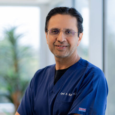
Meet your vein specialist
Dr. Syed Raza, MD
Vascular & Interventional Radiology · Founder, Premier Vein & Vascular Center
Fellowship-trained interventional radiologist certified by the American Board of Radiology and the Royal College of Radiologists (UK). Former Chief of Interventional Radiology at UTMB Galveston and Memorial Hermann Memorial City, with 15+ years treating varicose veins, venous reflux and peripheral arterial disease.
Leg Vein Anatomy
The leg vein system is a complex network that returns blood from the lower extremities to the heart. It comprises three primary types of veins, each with a unique function and location within the leg.
Superficial veins located just beneath the skin’s surface are easily visible when they become enlarged or twisted. The two main superficial veins include the great saphenous vein (GSV) and the small saphenous vein (SSV).
The GSV is the longest vein in the body, running from the foot to the groin. It’s also the most likely to become a varicose vein. The SSV runs along the back of the calf, from the ankle to the back of the knee. Like the GSV, it can also be a site of varicose vein formation.
Deep veins are located within your leg muscles. They carry the majority of blood back to the heart. The primary deep veins in the leg include:
- The anterior tibial vein running along the front of the lower leg
- The posterior tibial vein situated at the back of the lower leg
- The peroneal vein running along the side of the lower leg
- The popliteal vein located at the back of the knee
- The femoral vein running through the thigh
Perforator veins connect the superficial and deep veins, allowing blood to flow from the skin’s surface to deep within your leg muscles. Perforator veins are crucial for maintaining proper venous blood flow and preventing blood from pooling in the lower extremities.
Leg veins must fight gravity to circulate blood back to the heart. This is possible thanks to one-way valves that prevent blood from flowing in reverse. When these valves function properly, they efficiently pump blood upward. However, if these valves fail or weaken, blood can pool in the legs, leading to varicose veins.
Causes of Varicose Veins
Several factors increase the risk of developing varicose veins, including age, genetics, pregnancy, obesity, and prolonged periods of standing or sitting. As venous valve function weakens, blood flow becomes sluggish, and pressure builds in the veins. This increased pressure causes the veins to bulge and twist, resulting in the distinctive appearance of varicose veins.
It’s important to seek medical attention for varicose veins, as severe complications can occur if left untreated, including venous insufficiency, leg ulcers, and deep vein thrombosis. Premier Vein & Vascular Center in Katy, TX, specializes in diagnosing and treating vein disorders to help patients in the Houston area achieve healthier, more comfortable legs.

Medical Services for Varicose Veins
Several minimally invasive procedures can treat varicose veins, providing patients with safe and effective options tailored to their needs. Some of the cutting-edge vein treatments available at Premier Vein & Vascular Center include:
- Endovenous thermal ablation: This minimally invasive procedure uses heat energy to close affected veins, rerouting blood to healthier veins. Endovenous thermal ablation is safe and effective and requires minimal recovery time.
- Ambulatory phlebectomy: This surgical procedure involves removing varicose veins through tiny incisions. The technique is ideal for treating larger, more prominent varicose veins and results in minimal scarring and downtime.
- Sclerotherapy: This non-surgical treatment involves injecting a chemical solution into the affected veins, causing them to collapse and be absorbed by the body. Sclerotherapy is a particularly effective treatment for smaller varicose veins and spider veins.
- Non-thermal vein ablation: This minimally invasive treatment uses a medical adhesive or device to close the malfunctioning veins without heat. This option is ideal for patients not well suited for thermal ablation.
These varicose vein treatments offer minimal discomfort, short recovery time, and improved leg appearance. The specialists at our vein clinic will work with you to determine the best course of action based on the severity of your condition and medical history.
Contact Premier Vein & Vascular Center
Understanding the anatomy of leg veins is essential for maintaining good vascular health. By identifying the causes and contributing factors of varicose veins, our team can effectively address your venous disorders and help you fall back in love with your legs. We’re confident our minimally invasive procedures will relieve your discomfort and improve the appearance of your varicose veins. Don’t put off varicose vein treatment any longer! Our vein center in Houston is committed to providing expert care using the latest technologies available. Please call us at [company-phone id=2] to schedule a consultation, and we’ll discuss your needs as you start your journey toward a healthier, more confident future.
Leg Vein Anatomy in Motion: The Calf Muscle Pump
Valves alone cannot move blood uphill. The real engine of the leg’s venous return is the calf muscle pump. Every time you take a step, the calf muscles contract and squeeze the deep veins running through them, pushing blood upward toward the heart. When the muscles relax, the valves close to stop blood falling back, and the deep veins refill from the superficial veins through the perforators. Doctors sometimes call the calf the body’s second heart.
This is why the anatomy matters so much in daily life. Long periods of standing still or sitting with bent knees switch the pump off, so pressure in the ankle veins stays high. A stiff ankle, weak calf muscles or excess weight make the pump less efficient. And when valves fail, each step pushes blood up only for some of it to fall straight back down, so the pressure near the ankle never drops properly. That persistent high pressure, called venous hypertension, is what drives aching, swelling, skin changes and, eventually, ulcers.
Where Varicose Veins Usually Start: Junctions and Branches
Reflux in the superficial system usually begins at a few predictable points. Knowing where it starts is what allows us to treat the cause rather than just the visible bulges.
| Anatomical site | Where it is | What failure often looks like |
|---|---|---|
| Saphenofemoral junction | Groin, where the great saphenous vein joins the femoral vein | Varicose veins along the inner thigh and calf; aching and heaviness |
| Anterior accessory saphenous vein | Front of the thigh, running parallel to the great saphenous vein | Varicose veins on the front and outer thigh |
| Saphenopopliteal junction | Behind the knee, where the small saphenous vein joins the popliteal vein | Varicose veins on the back of the calf; ankle swelling |
| Perforator veins | Mainly the inner lower leg and ankle | Skin darkening, hardening and ulcers above the inner ankle |
| Pelvic escape points | Groin and upper inner thigh | Unusual varicose veins on the vulva, buttock or back of the thigh |
One leg can have more than one source. A full ultrasound map shows every segment that is refluxing so none is missed, which is a major reason varicose veins sometimes recur after treatment that addressed only part of the problem.
Primary vs. Secondary Vein Disease
Most varicose veins are primary: the vein walls and valves weaken over time, usually with a strong family tendency, and the problem stays in the superficial system. A smaller group of patients have secondary vein disease, where the trouble began in the deep veins. A previous deep vein thrombosis can scar deep vein valves, and a narrowing of a large pelvic vein, such as compression of the left iliac vein, can raise pressure in the whole leg.
The distinction matters because secondary disease may need a different treatment plan, and the deep veins must be assessed before any superficial vein is closed. A history of clots, swelling of the whole leg, or symptoms mostly in the left leg are clues we look for.
How pregnancy and aging change the picture
Pregnancy raises blood volume, relaxes vein walls through hormonal effects and adds pressure on the pelvic veins from the growing uterus, all of which strain the leg valves. Many women first notice varicose veins in pregnancy, and some improve after delivery while others persist. With age, vein walls gradually lose elasticity and valves can stretch apart so their leaflets no longer meet. Neither change is anyone’s fault, but both explain why vein disease so often becomes more noticeable over time and why periodic checkups make sense for people with a strong family history.
How We Evaluate Your Leg Vein Anatomy
- Focused history. We ask where your symptoms are, when they are worst, how long the veins have been present, and about previous clots, pregnancies, vein procedures and family history.
- Examination. We examine both legs standing, noting where varicose veins run, whether there is swelling, and any skin changes near the ankles. The pattern often hints at which junction or branch is responsible.
- Venous duplex ultrasound mapping. In our IAC-accredited vascular lab, the scan is performed with you standing or tilted so the veins fill as they do in daily life. Each junction, the saphenous veins, accessory branches and perforators are checked for reflux, and the deep veins are checked for old or new clot. You can read about this test on our venous ultrasound page.
- A clear plan. We show you which veins are failing and explain which treatment fits each part of your anatomy, in what order, and what recovery involves.
Matching the Varicose Vein Treatment to the Anatomy
Modern vein care is anatomy-driven. Rather than one treatment for every leg, we choose the tool that suits each faulty vein:
- Straight, refluxing saphenous or accessory veins are usually closed from the inside with endovenous thermal ablation or non-thermal ablation, especially when the vein lies close to a nerve.
- Twisting branch varicose veins near the surface are removed with ambulatory phlebectomy or treated with foam.
- Refluxing perforator veins near a skin change or ulcer can be closed with ultrasound-guided foam sclerotherapy or a small ablation catheter.
- Spider and reticular veins are treated with sclerotherapy once any underlying reflux is addressed.
Treating the source first and the branches second generally gives the most durable result.
Seek urgent medical care if you notice:
- Sudden swelling, pain and warmth in one calf or thigh
- Chest pain, sudden shortness of breath or coughing up blood
- A varicose vein that bleeds and does not stop with firm pressure and leg elevation
- A hot, red, hard and very painful vein with fever
When to See a Vein Specialist in Houston About Varicose Veins
If your legs ache, feel heavy or swell by evening, if varicose veins are enlarging, or if you notice skin darkening around the ankles, an ultrasound map of your leg veins is the logical first step. It explains why the veins formed and which treatment will address the cause.
Dr. Syed Raza is a fellowship-trained interventional radiologist with more than 15 years of experience treating varicose veins and venous reflux. If you are looking for a vein doctor in Houston or Katy, you can learn more on our varicose vein treatment page or call (832) 321-5355. Same-week appointments are usually available, and most insurance plans, including Medicare, are accepted.
Talk With a Specialist at Premier Vein
Have questions about your symptoms or treatment options? Dr. Syed Raza and the team at Premier Vein & Vascular Center can evaluate your concerns and explain your options clearly. Same-week appointments are usually available, and most insurance plans are accepted.
Frequently Asked Questions
Which leg vein is most commonly affected by varicose veins?
The great saphenous vein, which runs along the inner leg from the ankle to the groin, is the vein most often responsible for varicose veins. Reflux usually starts at its junction with the deep femoral vein in the groin.
Is it safe to close a saphenous vein?
Yes. A refluxing saphenous vein is already sending blood the wrong way, and most of the leg’s blood returns through the deep veins. After the faulty vein is closed, blood reroutes through healthier veins, which usually improves circulation.
What do perforator veins do?
Perforator veins connect the superficial veins under the skin to the deep veins in the muscles, and their valves let blood flow only inward toward the deep system. When they fail, high pressure is transmitted to the skin of the lower leg, which can contribute to discoloration and ulcers.
Why are varicose veins more common in the legs than the arms?
Leg veins must return blood against gravity over a long distance, so their valves face much higher pressure when you stand. Arm veins carry blood a shorter distance with less gravitational load, so valve failure there is uncommon.
Can varicose veins affect the deep veins?
Varicose veins themselves are in the superficial system, but superficial and deep veins are connected. Long-standing superficial reflux can be associated with deep vein problems, and a past deep vein clot can cause secondary varicose veins, which is why ultrasound checks both systems.
How does walking help leg veins?
Walking contracts the calf muscles, which squeeze the deep veins and push blood toward the heart. Regular walking lowers pressure in the ankle veins and is one of the simplest ways to ease aching and swelling from vein disease.
Do varicose veins on the back of the calf have a different cause?
Often, yes. Varicose veins at the back of the calf are frequently fed by the small saphenous vein, which joins the deep popliteal vein behind the knee. An ultrasound confirms the source so the right vein is treated.
Trusted Health Resources
- Chronic Venous Insufficiency, Society for Vascular Surgery
- Venous Insufficiency, MedlinePlus Medical Encyclopedia (NIH)
- Varicose Veins and Spider Veins, MedlinePlus (NIH)